Scientific Calendar February 2023
EDTA-dependent phagocytosis of platelets due to an underlying infection
Which measurement signal can indicate activation of neutrophils (NEUT-RI)?
Side fluorescence light (SFL)
Side-scattered light (SSC)
Peak height in PLT histogram
Forward-scattered light (FSC)
Congratulations!
That's the correct answer!
Sorry! That´s not completely correct!
Please try again
Sorry! That's not the correct answer!
Please try again
Notice
Please select at least one answer
Scientific background
The strategies of neutrophils within the immune response
A bacterial infection leads to an inflammatory condition in the human body; the innate immune system is triggered.
In the early phase of the innate immune response, neutrophils are activated and provide a first, non-specific line of defence against the pathogens. Doing so, they use two different strategies [1]. One of the strategies is secretion of proinflammatory cytokines and chemokines, which attract other neutrophils and activate other responses from the host towards the infection [2].
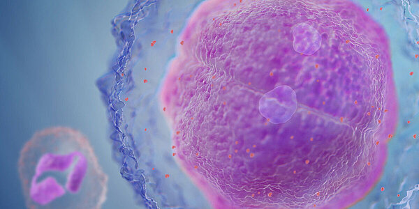
Additionally, neutrophils remove pathogens by phagocytosis and identify antigens to activate the adapted immune response [1]. During this phase the neutrophils are highly activated and their granularity and metabolic activity increase. This increase in staining density and number of granules in the neutrophil is called ‘toxic granulation’. Another indication for activation of neutrophils is the presence of vacuoles in the cytoplasm, which occur during increased phagocytic activity [3]. Activated neutrophils also present with increased metabolic activity in the cytoplasm, due to the production of chemokines and cytokines.
A further mechanism observed in activated neutrophils is the generation of neutrophil extracellular traps (NETs), which have been described as web-like structures composed of DNA, histones, and granules, with the purpose to entrap and eliminate pathogens [4].
Measuring elevated neutrophil reactivity and granularity on haematology analysers
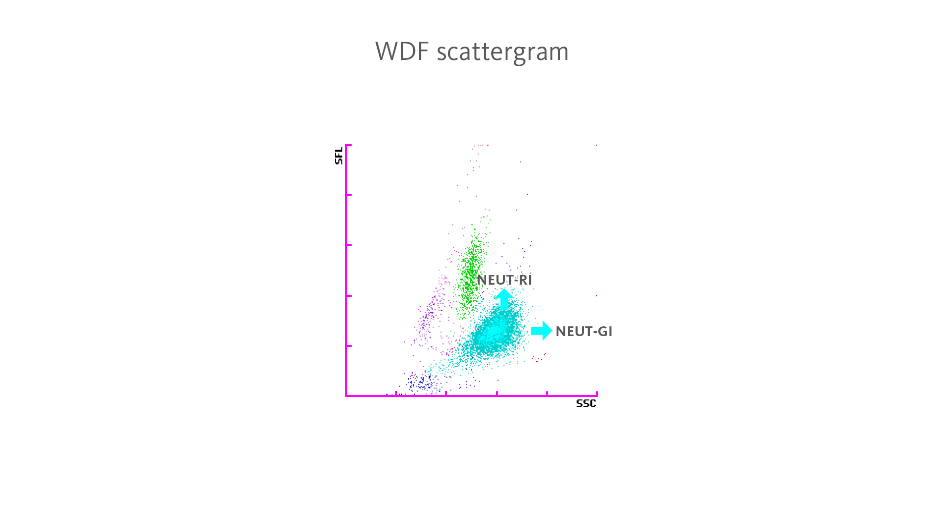
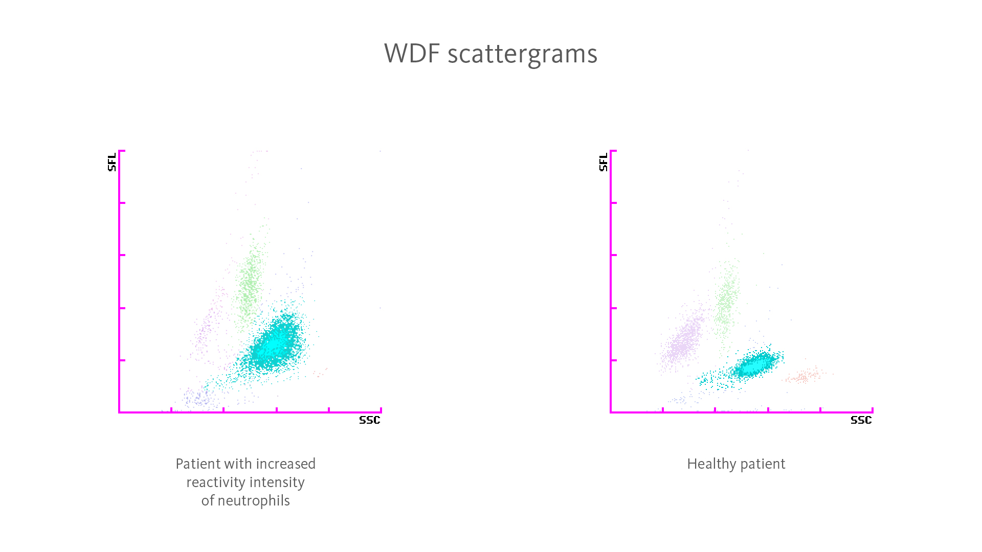
On XN-Series haematology analysers, a higher reactivity of neutrophils leads to an increased fluorescence intensity (SFL) in the WDF measurement channel, while an increase in granularity leads to higher scatter intensity (SSC).
With the ‘Extended Inflammation Parameters’ NEUT-RI (reactivity intensity of neutrophils) and NEUT-GI (granularity intensity of neutrophils) – which are diagnostic parameters – this becomes measurable.
Studies have shown that an increase in NEUT-RI and NEUT-GI is specifically present in bacterial infections [5,6].
The behaviour and fate of platelets in certain underlying conditions
In very rare cases, underlying conditions such as bacterial infections or oncological diseases can trigger autoantibodies against platelets [7]. These occur naturally in approximately 0.1% of the general population and up to 0.21% of hospitalised patients [8].
The anticoagulant EDTA has a chelating effect on the platelet membranes. By removing the Ca2+ ions it causes a confirmational change, exposing cryptic antigens on the GpIIbIIIa receptors on the platelet membranes. The autoantibodies now bind to the exposed antigens, which activate pathways leading to platelet agglutination, satellitism, and phagocytosis by neutrophils and, more rarely, monocytes [9–11].
This process called ‘EDTA-dependent pseudothrombocytopenia’ is not a disease but an in vitro phenomenon. Nevertheless, its identification is crucial to avoid unnecessary treatment and medication. Since the presence of pseudothrombocytopenia is often associated with autoimmune disorders or infections, it should therefore be followed up by investigation of possible underlying diseases [9]. Pseudothrombocytopenias caused by citrate and heparin are extremely rare [12].
In this case, the EDTA-dependent pseudothrombocytopenia was likely triggered by the Klebsiella pneumoniae infection.
Result interpretation
A patient presented at the hospital, complaining about malaise and fever with vomiting and symptoms of a urinary tract infection or pyelonephritis. An infection with Klebsiella pneumoniae was confirmed by blood culture later.
The blood count results obtained with an XN-Series analyser showed normal results for white blood cells, red blood cells, and slight thrombocytopenia. An increase in the Extended Inflammation Parameter NEUT-RI (NEUT-RI 65.0 FI; reference intervals 42.0–50.6 FI [13]) was observed. The flag ʽPLT Clumps?’ was also triggered.
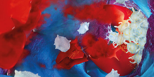
Digital imaging of the blood smear, taken from an EDTA blood sample, revealed strong activation of neutrophils, as well as platelet satellitism and platelet phagocytosis by neutrophils and monocytes (see Fig. 3). A smear of another blood sample taken 14 hours later still showed satellitism and phagocytosis, as well as vacuolation and destruction of neutrophils.
A new blood sample was drawn using citrate as anticoagulant which showed neither satellitism nor phagocytosis.*

*) The use of citrate anticoagulant is not supported by Sysmex and the possible impact on analytical performance needs to be validated by the user.
References
[1] Wright HL et al. (2010): Neutrophil function in inflammation and inflammatory diseases. Rheumatology. 49(9): 1618–31.
[2] Guerra FE et al. (2017): Epic Immune Battles of History: Neutrophils vs. Staphylococcus aureus. Front. Cell. Infect. Microbiol. 7: 286
[3] Zonneveld R et al. (2016): Analyzing Neutrophil Morphology, Mechanics and Motility in Sepsis: Options and Challenges for Novel Bedside Technologies. Crit Care Med. 44: 218–28.
[4] Brinkmann V et al. (2004): Neutrophil extracellular traps kill bacteria. Science. 303: 1532–5.
[5] Henriot I et al. (2016): New parameters on the hematology analyzer XN-10 (Sysmex™) allow to distinguish childhood bacterial and viral infections. Int J Lab Hematol. 39(1): 14–20.
[6] Cornet E et al. (2015): ): Contribution of the new XN-1000 parameters NEUT-RI and NEUT-WY for managing patients with immature granulocytes. Int J Lab Hematol. 37(5): e123–6.
[7] Sousa SM et al. (2020): Pseudothrombocytopenia: a case of platelet satellitism and phagocytosis by neutrophils. Platelets. 31(4): 541–3.
[8] Bartels PC et al. (1997): Screening for EDTA dependent deviations in platelet counts and abnormalities in platelet distribution histograms in pseudothrombocytopenia. Scand J Clin Lab Invest. 57(7): 629–36.
[9] Lippi G et al. (2012): EDTA-dependent pseudothrombocytopenia: further insights and recommendations for prevention of a clinically threatening artifact. Clin Chem Lab Med. 50(8): 1281–5.
[10] Tan GC et al. (2016): Pseudothrombocytopenia due to Platelet Clumping: A Case Report and Brief Review of the Literature. Case Rep Hematol. 2016:3036476.
[11] Ravel R et al. (1974): Platelet Satellitosis and Phagocytosis by Leukocytes. Lab Med. 5(6): 41–2.
[12] Sahin C et al. (2014): EDTA-induced pseudothrombocytopenia in association with bladder cancer. BMJ Case Rep. Bcr-2014-205130.
[13] L van Pelt J et al. (2022): Reference intervals for Sysmex XN hematological parameters as assessed in the Dutch Lifelines cohort. Clin Chem Lab Med. 60(6): 907–20.